Go back to the list of case studies .png) Click on the pictures to magnify and display the legends
Click on the pictures to magnify and display the legends| Case number: | 037 |
| Age: | 50 |
| Clinical presentation: | Postmenopausal woman with average risk of developing breast cancer presented with a left breast lump noticed 6 months ago. This was associated with pain at the site. Examination revealed a tender firm mobile lump less than a centimetre in diameter in the upper outer quadrant of the left breast. |
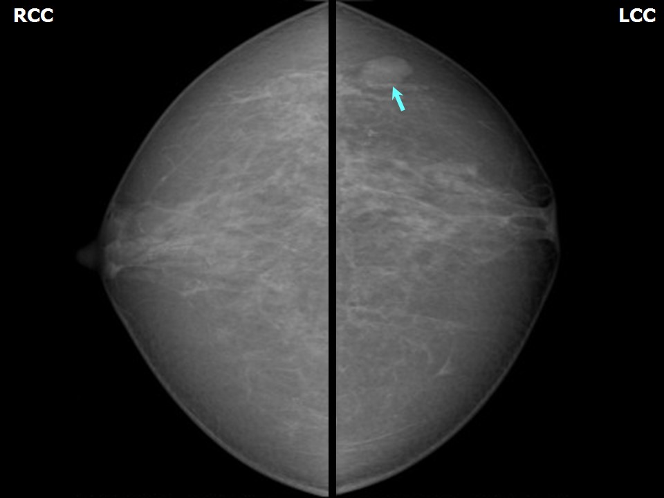
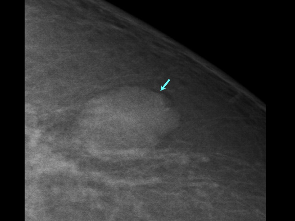
Mammography:
| Breast composition: | ACR category b (there are scattered areas of fibroglandular density) |
Mammography features: | |
|
| ‣ Location of the lesion: | Left breast, upper outer quadrant at 3 oclock, anterior and middle thirds |
| ‣ Mass: | |
| • Number: | 1 |
| • Size: | 1.5 × 1.0 cm |
| • Shape: | Irregular |
| • Margins: | Microlobulated |
| • Density: | Equal |
| ‣ Calcifications: | |
| • Typically benign: | None |
| • Suspicious: | None |
| • Distribution: | None |
| ‣ Architectural distortion: | None |
| ‣ Asymmetry: | None |
| ‣ Intramammary node: | None |
| ‣ Skin lesion: | None |
| ‣ Solitary dilated duct: | None |
| ‣ Associated features: | None |
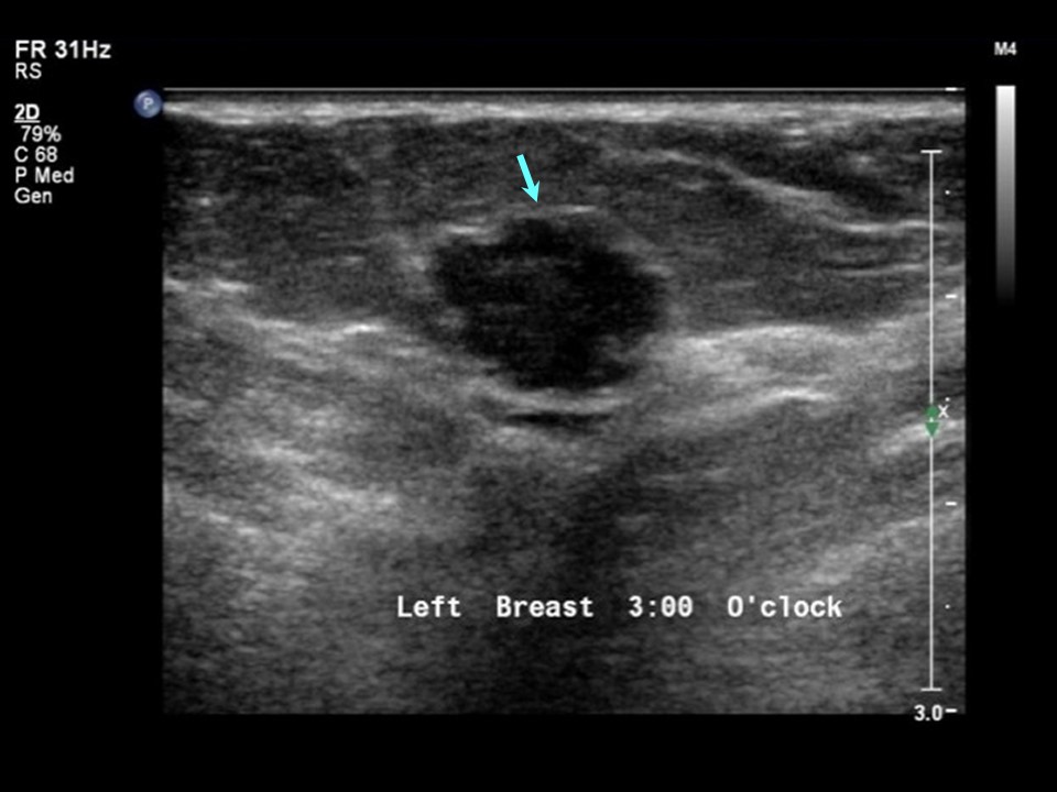
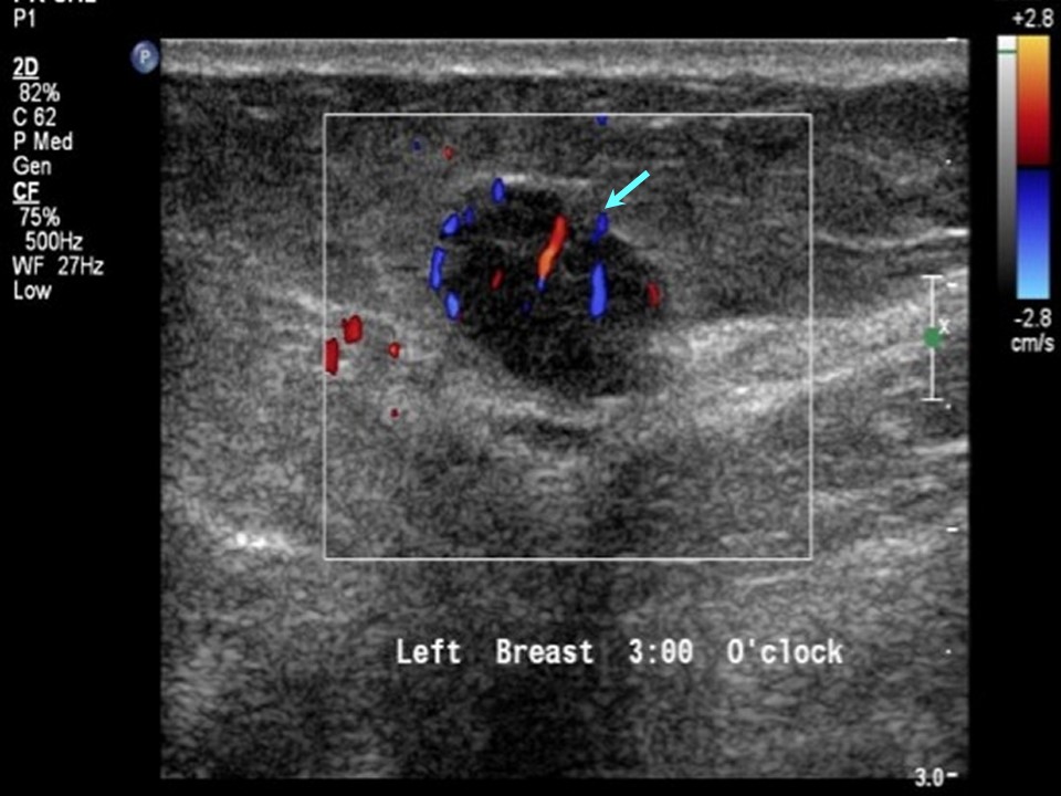
Ultrasound:
| Ultrasound features: Left breast, upper outer quadrant at 3 oclock |
|
| ‣ Mass | |
| • Location: | Left breast, upper outer quadrant at 3 oclock |
| • Number: | 1 |
| • Size: | 1.0 cm in greatest dimension |
| • Shape: | Irregular |
| • Orientation: | Not parallel |
| • Margins: | Microlobulated |
| • Echo pattern: | Hypoechoic |
| • Posterior features: | No posterior features |
| ‣ Calcifications: | None |
| ‣ Associated features: | Internal vascularity |
| ‣ Special cases: | None |
BI-RADS:
BI-RADS Category: 4B (moderate suspicion of malignancy)
Further assessment:
Further assessment advised: Referral for cytology and for excision biopsy
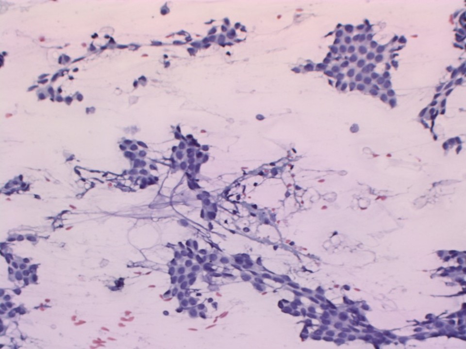
Cytology:
| Cytology features: | |
|
| ‣ Type of sample: | FNAC |
| ‣ Site of biopsy: | |
| • Laterality: | Left |
| • Quadrant: | Outer lower |
| • Localization technique: | Palpation, rubbery consistency felt through the needle |
| • Nature of aspirate: | whitish |
| ‣ Cytological description: | Cellular smear showing elongated, branching epithelial fragments of regularly arranged cohesive cells. Single, bare bipolar or oval nuclei are scattered in the background. Loose fibromyxoid stroma are also seen |
| ‣ Reporting category: | Benign |
| ‣ Diagnosis: | Fibroadenoma |
| ‣ Comments: | None |
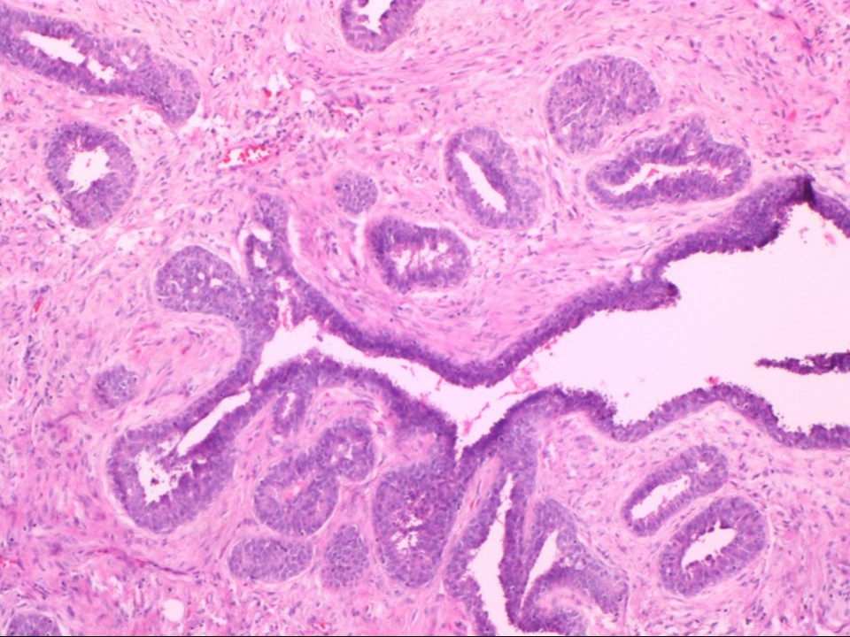
Histopathology:
Lumpectomy
| Histopathology features: | |
|
| ‣ Specimen type: | Lumpectomy |
| ‣ Laterality: | Left |
| ‣ Macroscopy: | Lumpectomy specimen (9.0 × 7.0 × 3.0 cm) with skin flap. Cut surface shows a firm white area (1.2 × 1.0 × 0.5 cm), 1.0 cm from the inferior margin below the skin. Another whitish area (3.0 × 2.0 cm) is seen superomedially |
| ‣ Histological type: | Sections from lumpectomy specimen reveal histological features of a fibroadenoma having intracanalicular and pericanalicular patterns. A few ducts in the fibroadenoma show features of UDH. The overlying skin is unremarkable. The adjacent fibroadipose tissue shows foci of fibrosis |
| ‣ Histological grade: | |
| ‣ Mitosis: | |
| ‣ Maximum invasive tumour size: | |
| ‣ Lymph node status: | |
| ‣ Peritumoural lymphovascular invasion: | |
| ‣ DCIS/EIC: | |
| ‣ Margins: | |
| ‣ Pathological stage: | |
| ‣ Biomarkers: | |
| ‣ Comments: | |
Case summary:
| Postmenopausal woman presented with left breast lump. Diagnosed as mass of suspicious morphology, BI-RADS 4B on imaging and as fibroadenoma left breast on cytology. Fibroadenoma showed foci of epithelial hyperplasia on histopathology. |
Learning points:
- Fibroadenoma can reveal atypical features on ultrasound suspicious of breast carcinoma, such as irregular shape, heteroechogenicity, microcalcifications within the mass, or posterior shadowing. Microlobulated margins are a feature of atypical fibroadenoma. These atypical appearances are caused by interdigitation of the surrounding parenchyma.
|